Doppler Disasters

Ultrasound of the Abdominal Aorta

measurementaoThe aorta; by far one of my favorite great vessels to lay my sono-eyes on–sure, sure, it can be challenging on occasion but hey–   “A challenge only becomes an obstacle when you bow to it.” ― Ray Davis

That said, there are sono-standards published by the AIUM which I will paste below; this gives an overview of what is to be included in an aortic ultrasound, but leaves a bit to be desired when talking technique; cue the sono-insight from the girl at the disposal of sono-tip seekers. At your service 😉

SAGITTAL IMAGING
When you’re scanning the aorta in sagittal, you will be obtaining AP measurements at the proximal, mid, and distal segments. The most frequent uncertainty of students–and some rads–yeah, they don’t know EVERYTHING–just don’t tell them that ;)–is where to place the calipers.

The aorta is measured from outside wall to outside wall according to AIUM standards.

It’s also a nice touch for reference, to use other vessels as landmarks along the length of the aorta when possible; this further illustrates your location for the radiologists. At the proximal segment, I will usually try to get the SMA origin. While there’s not much else, in terms of vessels arising from the anterior of the abdominal aorta, that you can ROUTINELY use as a reference point in sagittal; if you’re feeling squirrely you can try for the IMA (distal to the SMA).
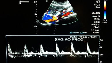
I also drop color and Doppler to get a pretty waveform if possible in each segment because I like to and I can, though most wouldn’t consider it “necessary” in a normal abdominal aorta. However; you may find that doing what isnt necessary, yet still relevant can be a way of branding yourself;though not all rads will give you accolades for it.

Now, if during your sagittal evaluation, you run into an aneurysm, you will want to get the craniocaudal length (i.e. where it starts to where it ends). You also need to capture and document any intraluminal pathology such as thrombus, calcific plaque, or dissection. Also be sure to drop color in the aneurysm  to demonstrate the disturbance of flow within the greatest area of dilatation; this will also highlight areas without flow, such as mural thrombus and/or dissection, where the absence of color can indicate a false lumen.   You will continue the same technique through the bifurcation and bilaterally into the proximal iliacs to get the AP dimension.

aortasagproxaortasagcolorprox

aortasagdoppprox


TRANSVERSE
Your transverse imaging will mirror the protocol above in the axial plane. Take your TRV measurements at the proximal (celiac trunk as a landmark), mid (renal vessels landmark) distal, and the proximal iliacs where it bifurcates. I typically drop color on the trv iliacs to highlight the vessels since they are so small and can easily be lost in the bowel gas.


trvaortamid

trvaormidcolor


AIUM: Specifics of the Aorta Sonography Examination

At a minimum, the diagnostic examination includes the following, when feasible:

  • Longitudinal images of the proximal, mid, and distal abdominal aorta with anterior to posterior measurement
  • Transverse images of the proximal, mid, and distal abdominal aorta
  • Longitudinal images of the right and left common iliac arteries with anterior to posterior measurement
  • Transverse image of the right and left common iliac arteries near the aortic bifurcation
  • Color Doppler imaging and/or spectral Doppler imaging with waveform analysis, as indicated.
  • Documentation of mural thrombus if present

(more…)

Portal Vein Pains: Turn your Doppler Woes into Doppler WOWS

ultrasound tips & tricks

Now the portal vein is something with which you should become very familiar– not just because you see it every time you scan an abdomen, but it is also one of the most commonly ordered vascular Doppler studies ordered for radiology ultrasound–so make it a point to get to know it now and love it later.

To best understand portal venous flow it is first important to understand what is normally occurring in the abdomen so that you understand how common pathology will affect it.

The portal vein is a result of the joining of veins from two areas in the body–the mesenteric veins and the splenic vein —these veins direct all the blood flow from the spleen and bowels into the liver to be cleaned and to harvest nutrients.

So first stop and picture this–blood from the bowels travels upwards/superiorly towards the head of the pancreas, and blood from…

View original post 504 more words

How to find and recognize the SMA by Ultrasound for sonography students

Superior Mesenteric Artery;  Go from SMApprehensive to SMAwesome

The Mesenteric Arteries are often requested by physicians to be interrogated with Doppler to rule out abnormalities in flow that could contribute to bowel conditions such as ischemia. I also include it in any Doppler request, even if its only the hepatic vessels being requested–just because I love it. You should try to love it too. It makes you more valuable to Radiologists and to techs who loathe the little blood bearing beasts. So lets start with the SMA.

The SMA is one of the easiest arteries to find by ultrasound really, especially once you become familiar with the waveform which is VERY high resistance (in an NPO state). Place your probe up by the xyphoid process, where you would place it to get the left lobe of the liver.

You’ll notice the aorta rising up towards your probe (aka anteriorly). Turn your color on–the SMA has very prominent flow and turbulent appearance at its origin–and its easy to distinguish from the celiac–which will come into play as the first branch off of the abdominal aorta–because remember that the SMA runs sagittal–along WITH the aorta–if you can find the aorta, you’ll also have the SMA in view. Its only about 1 cm from the celiac axis, and its velocity is much greater than the aorta–SMA is around 200-300 cm/s with a more narrow lumen-the aorta will be under 100 cm/s and more visibly pulsatile.

The waveform for the SMA is VERY distinct. This is what normal looks like.

Abnormal:notice how very sharp and peaky the waveform is in systole, diastolic velocity is elevated and there’s no dip below baseline as you see in the normal waveform, the peak systolic velocities are in excess of 300 cm/s with ischemic disease/stenosis.


So, there you go, you’re an official SMA waveform interpreter…. bet you cant wait ’til your next NPO patient comes in for a simple gb ultrasound…I know you’re gonna slide on over and check out the mesenteric arteries…mmm hmm, just make sure you turn down the volume on your PW dial so you don’t freak the patient out (or find yourself at the end of a patient’s insatiable appetite for answers!). Good luck, SMA sleuth!

Portal Vein Pains: Turn your Doppler Woes into Doppler WOWS

Now the portal vein is something with which you should become very familiar– not just because you see it every time you scan an abdomen, but it is also one of the most commonly ordered vascular Doppler studies ordered for radiology ultrasound–so make it a point to get to know it now and love it later.

To best understand portal venous flow it is first important to understand what is normally occurring in the abdomen so that you understand how common pathology will affect it.

The portal vein is a result of the joining of veins from two areas in the body–the mesenteric veins and the splenic vein —these veins direct all the blood flow from the spleen and bowels into the liver to be cleaned and to harvest nutrients.

So first stop and picture this–blood from the bowels travels upwards/superiorly towards the head of the pancreas, and blood from the spleen travels from left to right towards the pancreatic head where it becomes the portal vein.

So, by this diagram, you should be able to picture how the blood should flow based on where you place your probe. I absolutely always recommend utilizing an intercostal approach to assess the direction of flow–you can almost always visualize the portal vein on anyone despite body habitus/obstacles common to ultrasound. This allows you to become familiar with what you should see every time. RED flow–intercostally, the flow will be advancing towards the transducer so normal should be red. Normal is also called hepatopedal flow—-whereas abnormal is called hepatofugal flow–now you may not be exposed enough to be able to readily identify flow terminology–so here’s the trick I used in school, and still use by the way: hepatofugal is bad–its reversal of flow caused by a resistive force that prevents entry of blood into the liver–hepatofugal is as bad as the f-word. We’ll call it ‘fudge’ as a tribute to the  movie ” A Christmas Story” that I absolutely love for exactly 28 days commencing immediately post-Thanksgiving day until the 25th of December–no later. I digress. So I think in my head hepatofudgal. I know less than academic, but it works. No ‘fudging’-fail method of remembering the direction of hepatic flow.

Portal vein directional flow


Now, when a patient has cirrhosis, the liver becomes tough like a bad steak that’s hard to get your teeth through–same for blood through the liver–the blood doesn’t want to slow its roll, so it keeps on coming, but has nowhere to go, so it backs up into the vessels it came from.. Think of a panty hose on a faucet that people like my parents used to use to catch lint from the washer–when the hose becomes full of lint, its like a cirrhotic liver–and what happens (I know because I used to watch it) the hose becomes dilated like a water balloon as the water backs up, and then it comes out of whatever place gives the least resistance.. So in the case of cirrhosis, it will back up into the portal vein first–and you’ll notice its dilated (2 cm is upper of norm) when you turn on the color, it will be EITHER blue, OR bidirectional–(bidirectional is a turbulent mix of red and blue and indicates early fibrotic changes–and it will reflect in the waveform as well.)


Portal Hypertensive Waveform

Turbulence in Portal Vein


And then of course, you will look at other areas that will hold the overflow, refer back to the picture, where will it back up into?


Splenic varices with PHTN

Spleen-it will be big and bloated with blood–bigger than 12 cm and typically have varices from the increased pressure and volume of blood backing up from the liver


Umbilical vein (remnant reopens under pressure) you’ll see it as a weird vessel smack dab    in the middle of the left lobe of the liver.

And now you got it, right?


You Got This!!

You Got This!!