FacebookTwitterGoogle+LinkedInBloggerPinterestEmail

Welcome to the site…….at least you found SOMETHING…right? 😉

This is one of the venues I am posting the most sought after sonography tips and tricks of the trade; of course I will address any additional information you needy students may have; at this point I am certain you have, at a minimum, honed your skill of torturing the technologists with your tedious interrogations. Hope you all find it useful!

Please feel free to contact me directly with any questions or comments using the contact tab at the top and I will reply as soon as I possibly can in an effort to satisfy inquiring minds; I am at your service 😉

The ‘Down Under’ and Women’s Health (Down There)

It’s no longer a secret that health care providers in America are inadvertently threatening the health of women; while governing bodies such as the Center for Disease Control (CDC) and the Food and Drug Administration (FDA) are typically regarded as the authority on etiology of disease and best practices to prevent the spread of pathogens- they are not always as they seem. That is, the chemicals include in their recommendations for high level disinfection are not supported by rigorous scientific data and employing these chemicals for HLD processing of transvaginal probes places patients at risk for HPV exposure.

Even more disconcerting is that the both the FDA and CDC have been aware of this for more than a year.

While efforts to enact changes that protect the health of the American population have fallen on deaf ears at the CDC and FDA; Australia was listening.  In an effort to protect the health of women as well as the future and reputation of sonography, the scientists at Nanosonics ltd were proactive in their approach to ensure their own innovative automated technology for high level disinfection (Trophon EPR) was effective against HPV: an under-represented and growing threat to women’s health.  Here’s what they found:

australia

MUST READ – Important Information Concerning Human Papilloma Virus (HPV)

Nanosonics has been very busy working with customers around the world as we transform the ultrasound probe disinfection practices to fast, safe, automated, effective and environmentally friendly high level disinfection.

Part of our mission is to help protect people and with this in mind, we share new scientific information and evidence based best practices to help reduce HAIs.  Today we are faced with exceedingly important research results that must be shared with as many people as possible.  Distinguished Professor, Craig Meyers from Penn State College of Medicine, presented these research results last week at the SHEA conference in Orlando.

In summary, these results are:

1.       HPV is cancer causing – High-risk HPV accounts for 5% of all cancers worldwide and is responsible for almost all cases of cervical cancer, and is a leading cause of oral, throat, anal and genital cancers.

2.       Ultrasound exams pose a real HPV transmission risk – A number of studies have demonstrated the transmission risk of HPV from ultrasound probes as between 3% and 7% of probes used in trans-vaginal examinations were found to be positive to HPV even with the use of a sheath and post wiping.

3.       Until now no one has been able to grow the human, infectious virus to test against the FDA standards for disinfection – Prior to now there has not been an appropriate test to determine the effectiveness of disinfectants against the natural, infectious form of HPV. This new test has been developed at Penn State College of Medicine and Brigham Young University.

4.       HPV is not being killed with old methods – Studies using this new validated test have confirmed that OPA and glutaraldehyde are in fact ineffective in at killing HPV

5.       trophon EPR is effective against HPV – The new study presented at SHEA last week has reconfirmed that OPA is ineffective against high-risk, cancer causing HPV and, more importantly, proven that trophon EPR is totally effective in completely inactivating HPV.

We trust this information will be helpful as you prepare your facility and practices to effectively fight the potential transmission of HPV via ultrasound transducers. Please see the attached HPV Information Handout and there is additional information on www.hpvdisinfection.com.

.

Human Papillomavirus and Transvaginal Ultrasounds: Sonography Students May Be Exposed to More Than Humiliation

caduceus-scalesThe recent allegation brought about against Valencia College by two medical sonography students claim transvaginal sonographic examinations were a mandatory part of the program to which they were forcibly subjected.  Valencia’s response, “The use of volunteers — including fellow students — for medical sonography training is a nationally accepted practice. .”, resuscitates an ongoing issue rooted in the FDA’s deficient criteria in characterizing high level disinfectants (HLD).

Currently, the characterization of a chemical as an HLD is not specific to its use; in other words, they are NOT tested for the diseases to which they will most likely be exposed and employed to kill. In 2014, research conducted by Meyers et al, demonstrated the impotence of the 11 most commonly employed high level disinfectants against human papillomavirus-16 (HPV); the strain linked to cervical and oropharyngeal cancers [1]. Among the chemicals that were ineffective at deactivating HPV-16 were glutaraldehyde and ortho-phthalaldehyde (OPA). These chemicals are characterized by the FDA as high level disinfectants and recommended by the CDC [2] for HLD of semi critical items. As a result, they are widely utilized for HLD of transvaginal, transrectal, transesophageal probes, and flexible endoscopic probes; we know these chemicals by their product names: Cidex Plus, Cidex OPA, Metrocide etc.

This data has now been reinforced by newly published research by Craig Meyers from Penn State College of Medicine that demonstrates the impotence of some of the most common disinfectants used in healthcare today.

Sadly, the FDA and CDC are aware that the chemicals they recommend are inadequate and pose serious risk to patients, however the patients and students subject to this examination are not. In an era that emphasizes patient rights, the FDA and CDC fail to exemplify this purported priority.  Despite the growing research supporting glutaraldehyde is ineffective against HPV, and despite  diligent attempts to communicate this to the FDA, EPA and CDC; correspondence that includes a formal petition I filed with the FDA in 2014: they have yet to act in the interest of safety, disease prevention and basic human rights. The victims of the FDA and CDC’s failure to act have now expanded to include sonography students, such as the Valencia College students, who already feel victimized and humiliated and now face the grim reality that they may have also been forcibly exposed to an incurable cancer causing disease. For more information about HPV click here.

Saving One-Third of a Woman is Not Impressive to Payers

How a Study Design Can Prevent Early Detection of Breast Cancer

onethird2Published research influences healthcare-as it should; given the research contains a valid design: not limited to being bias-free, and reflecting inclusive consideration of external variables potentially impacting results. Every aspect of the design should withstand challenges from critics and colleagues; from sample population selection to outcome measures. Reported results from original research are critical because they have the potential to be instrumental in the decision making process of providers and payers; these decisions invariably affect healthcare. It plays a crucial role in defining treatment options/technology, access and reimbursement which will ultimately and profoundly impact the patient.

Below is my evaluation of the study by Sprague et al: Benefits, Harms, and Cost-Effectiveness of Supplemental Ultrasonography Screening for Women With Dense Breasts which was recently published by The Annals  of Internal  Medicine in response to the breast density inform law adopted by 19 states in the US.


 The Study

Background: Many states have laws requiring mammography facilities to tell women with dense breasts and a negative screening mammography result to discuss supplemental screening tests with their providers. The most readily available supplemental screening method is ultrasonography, but little is known about its effectiveness.
Objective: To evaluate the benefits, harms, and cost-effectiveness of supplemental ultrasonography screening for women with dense breasts.
Design: Comparative modeling with 3 validated simulation models.
Data Sources: Surveillance, Epidemiology, and End Results Program; Breast Cancer Surveillance Consortium; and medical literature.
Target Population: Contemporary cohort of women eligible for routine screening.
Time Horizon: Lifetime.
Perspective: Payer.
Intervention: Supplemental ultrasonography screening for women with dense breasts after a negative screening mammography result.
Outcome Measures: Breast cancer deaths averted, quality-adjusted life-years (QALYs) gained, biopsies recommended after a false-positive ultrasonography result, and costs.
Results of Base-Case Analysis: Supplemental ultrasonography screening after a negative mammography result for women aged 50 to 74 years with heterogeneously or extremely dense breasts averted 0.36 additional breast cancer deaths (range across models, 0.14 to 0.75 deaths), gained 1.7 QALYs (range, 0.9 to 4.7 QALYs), and resulted in 354 biopsy recommendations after a false-positive ultrasonography result (range, 345 to 421 recommendations) per 1000 women with dense breasts compared with biennial screening by mammography alone. The cost-effectiveness ratio was $325 000 per QALY gained (range, $112 000 to $766 000 per QALY gained). Supplemental ultrasonography screening for only women with extremely dense breasts cost $246 000 per QALY gained (range, $74 000 to $535 000 per QALY gained).
Results of Sensitivity Analysis: The conclusions were not sensitive to ultrasonography performance characteristics, screening frequency, or starting age.
Limitation: Provider costs for coordinating supplemental ultrasonography were not considered.
Conclusion: Supplemental ultrasonography screening for women with dense breasts would substantially increase costs while producing relatively small benefits.
Primary Funding Source: National Cancer Institute.


EVALUATION

Target Population: The study fails to include the age group (40-50 years) for whom ultrasound has been demonstrated as 13.7% more specific than mammography. [1] The study accounts for 40% of women (estimated percentage of women with dense breasts) between 50-74 years of age but does not include women aged 40-50. Since breast density decreases with age, it stands to reason that the group aged 40-50 years may have yielded an overall patient benefit more favorable to adjunct sonographic evaluation of dense breast tissue. The selected population woefully underrepresents a proportion of the population that has been shown to substantially benefit from breast sonography. Additionally, the inclusion of those aged 40-50 would have expanded the sample population by approximately 40% [2].

Methods: Evaluation of handheld sonography as an adjunct modality limits the veracity of reported results. Handheld sonography is notoriously operator-dependent; the widespread variance in technique, experience, and interpretation limits the validity of the study. Inclusion of automated breast ultrasound (ABUS), a more advanced form of breast sonography that eliminates these variables, may have returned a more promising report of patient benefit in lieu of the cost prohibitive conclusion that favors a payer’s objective.

Outcome Measures

Deaths Averted: The study fails to account for the impact of early detection/aversion of invasive diagnosis that is potentially achieved when utilizing sonography in conjunction with mammography. Approximately 78% of cancers are invasive at the time of diagnosis; invasive cancer treatment accounts for $12-16 billion dollars. The average cost per patient is 5 times greater when diagnosed at an advanced versus early/in situ stage. [3, 4] Currently deaths from breast cancer constitute approximately 10-13% of total diagnoses per year [3]. The outcome fails to assess the impact on 90% of breast cancer diagnoses. Why not measure the impact of early detection on cost, or the impact of adjunct use of breast sonography on reduction of invasive diagnoses? Either of these designs may have yielded a more impressive result than focusing primarily on death aversion.

Study Considerations: Existing factors that contribute to overall costs

Modality Accuracy: This study failed to consider current factors that contribute to the overall cost; factors that are mitigated by the addition of ultrasound as an adjunct modality to mammography in dense breast evaluation. False negative rates:mammography misses 20-28% percent of breast cancers that are present at the time of screening compared to 12.8% false negative rate of breast sonography [3]. Combined, mammography and breast sonography reduces overall multimodality screening to 4% false negative (ACRIN). False positive rate of breast sonography alone is 7.5% [5], compared to a false positive rate of 50-60% in mammography[6]

Interpretation Variance: The recommendation for breast biopsy may be exclusive of the modality in question. Instead, it may be specific to the training of the interpreting radiologist.  There are standards for breast imaging interpretation outlined by the ACR (BIRAD/ACR); not all practices are accredited and/or not all radiologists follow these guidelines when interpreting breast imaging, particularly sonographic imaging.  Perhaps the variance in training and interpretation of mammo/sonography is a causative factor in the number of subsequent biopsy recommendations. In addition to defensive medicine, perhaps the BIRADS guidelines should also be evaluated for classification and recommendations that may be outdated and/or contribute the the incidence of unnecessary biopsies.

Accuracy of Histology Reports. False negative rate of breast biopsies: 20-43% of biopsies are misdiagnosed during the pathological analysis of tissue specimens [7] . Training variance amongst pathologists, experience, equipment and expertise all impact the final diagnosis. This could have been explored as a potential contributor to “unnecessary biopsies” if the results were negative at the time of the study.

Summary

The accuracy of results are influenced by many factors, though expansion of the target population is warranted simply by the incidence of breast cancer in women over 40. Technologically, the incorporation of automated breast ultrasound (ABUS) as an adjunct to mammography in breast cancer detection in women with dense breasts, would likely yield greater accuracy and potentially reduce the false positives and ‘unnecessary biopsies’ attributed to handheld sonography. If the study was redesigned to include outcome measures such as detection of breast cancers earlier than they would have been diagnosed with mammography alone, the impact has the real potential to reflect a reduction in the percentage of invasive diagnoses as well as the associated cost of treatment. Another consideration is the impact that standardizing the interpretive process and tissue specimens may have on reducing costs.

When figures are reported fractionally, i.e. one third of a woman saved per thousand women–they reflect a seemingly paltry impact disproportionate to the price tag each whole woman costs (bear in mind that the study was performed from a payer’s perspective). However, when applied to the current population of females in the US, the results are a bit more human (though I can’t say the same of the payers). According to the US Census there are 76 million women in the US over age 40, 21 million are between 40-50 years, 55 million over 50. Not taking into consideration the limitations of the study and reported results, the overall impact of combining handheld ultrasound with mammography in women with dense breasts would be:

Deaths Averted/Lives Saved; 27,360 women, mothers, daughters, wives, sisters, friends, aunts, and/or grandmothers.

References
[1] Houssami N, Irwig L, Simpson JM, McKessar M, Blome S, Noakes J. Sydney Breast Imaging Accuracy Study: Comparative sensitivity and specificity of mammography and sonography in young women with symptoms. AJR Am J Roentgenol. 2003 Apr;180(4):935-40.
[2] US Census. http://www.census.gov
[3] Breast Cancer Facts and Figures 2013-2014. Cancer.org. http://www.cancer.org/acs/groups/content/@research/documents/document/acspc-042725.pdf
[4] Marj P. Zimmerman, MS, BSPharm, RPh; and Stanton R. Mehr Breast Cancer: Will Treatment Costs Outpace Effectiveness? AJMC: Evidence Based Oncology. 2012
[5] University of Virginia Health System: Cancer Center. Breast Ultrasound. 2014 http://cancer.uvahealth.com/patient-care/screening-and-genetics/breast-cancer-screening/breast-ultrasound
[6] Susan G Komen. Komen.org. Mammogram Accuracy. 2014.   http://ww5.komen.org/BreastCancer/AccuracyofMammograms.html#sthash.uf0YQl00.dpuf
[7] Susan G Komen for the Cure. 2006. Why Current Breast Pathology Practices Must Be Evaluated.  http://ww5.komen.org/uploadedFiles/Content_Binaries/PathologyWhitePaperB2.pdf
[8] Rotten D1, Levaillant JM. The value of ultrasonic examination to detect and diagnose breast carcinomas. Analysis of the results obtained in 125 tumors using radiographic and ultrasound mammography.Ultrasound Obstet Gynecol. 1992 May 1;2(3):203-14.
[9] Sprague, B et al. Benefits, Harms, and Cost-Effectiveness of Supplemental Ultrasonography Screening for Women With Dense Breasts ONLINE FIRST
Ann Intern Med. Published online 9 December 2014 doi:10.7326/M14-0692

Are You a Danger to YOUR Patients?

probetv

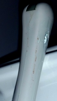
Post procedural transvaginal probe: contaminated surface despite the use of a probe cover.

Probe sheaths are often used with endocavity ultrasound transducers to prevent disease transmission between patients, however; studies have shown probe cover leakage of up to 81% [4,5,9,10,11]. For that reason, probe covers are required to be used in conjunction with high level disinfection (HLD) of endocavity probes.

But, what does it mean for a chemical to be termed a ‘high level disinfectant’ versus a ‘low level disinfectant’ or sterilant?

“ FDA defines a high level disinfectant as a sterilant used under the same contact conditions except for a shorter contact time. Therefore, products with high level disinfectant claims should first qualify as a sterilant by passing the Association of Official Analytical Chemists (AOAC) Sporicidal Test (Sporicidal Activity of Disinfectants, AOAC 6.3.05:1995, Official Method 966.04) ..”

You’d probably be pretty surprised to find out that the characterization of a chemical as an HLD is not specific to its use; in other words, they are NOT tested for the diseases to which they will most likely be exposed and employed to kill. Well, it shouldn’t have come as much of a surprise when earlier this year, research conducted by Meyers et al, demonstrated the impotence of the 11 most commonly employed high level disinfectants against human papillomavirus-16 (HPV); the strain linked to cervical and oropharyngeal cancers [1]. Among the chemicals that were ineffective at deactivating HPV-16 were glutaraldehyde and ortho-phthalaldehyde (OPA). These chemicals are characterized by the FDA as high level disinfectants and recommended by the CDC [2] for HLD of semi critical items. As a result, they are widely utilized for HLD of transvaginal, transrectal, transesophageal probes, and flexible endoscopic probes; we know these chemicals by their product names: Cidex Plus, Cidex OPA, Metrocide etc.

The alarming rate of probe cover failure nearly guarantees probe surface exposure to pathogens;combine that with an ineffective HLD and you’ve a recipe for diseased disaster and provider liability which necessitates immediate mitigation through HLD revision at the Federal Level.


probetv3 probetv2

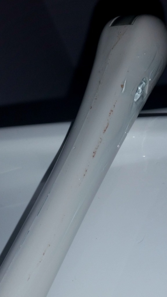
The images above show an endocavity transducer following a transvaginal examination I performed. The images were taken immediately after removing the probe cover- just prior to HLD.  Clearly, there is contamination of the probe surface that occurred despite the use of a barrier.  If I was using Cidex/Metrocide, and my patient was previously exposed to HPV (an estimated 25% of people) it would remain on the surface of the probe even after HLD. Its only logical to conclude that, since blood FROM the patient was able to penetrate the sheath and contaminate the surface of the probe, that any pathogens that survive the HLD process could also be transmitted in kind, TO the next patient. (Luckily, my organization was proactive and is using a vaporized Hydrogen Peroxide system which does kill HPV) we bought through GE at the time, however; I heard they can now be purchased directly from Nanosonics.

For those of you still only performing low level disinfection (LLD) on transvaginal probes–look again at the images above and imagine that probe being used on you after only being cleaned with a spray and/or wipes. LLD guarantees you are giving your patients more than an ultrasound examination; you are transmitting disease to your patients.

Sadly, the CDC is aware that the chemicals they recommend are inadequate and pose serious risk to patients. Their guidelines are the very basis upon which most health care organizations develop their infection control measures in an effort to ensure patient safety; making the CDC’s failure to react and revise their recommendations even more egregious. There alternatives to CIDEX PLUS and CIDEX OPA on the CDC’s list that can be instituted to process intracavity ultrasound probes. Great organizations shouldn’t have to be forced to take the most progressive approach in patient safety measures. The CDC might be taking their time, at the expense of patients’ health; you don’t have to.


References
[1]Ryndock E, Conway M, Meyers C, Robison R Meyers J, “Susceptibility of high-risk human papillomavirus type 16 to clinical disinfectants,” Susceptibility of high-risk human papillomavirus type 16 to clinical disinfectants, The journal of antimicrobial chemotherapy vol. 69, no. 6, pp. 1546-1550, 2014.
[2]Ph.D., M.P.H., David J. Weber, M.D., M.P.H., and the Healthcare Infection Control Practices Advisory Committee William A. Rutala, “Draft Guideline for Disinfection and Sterilization in Healthcare Facilities,” Atlanta, 2/20/2002.
[3]Casalegno J. et al, “High Risk HPV Contamination of Endocavity Vaginal Ultrasound Probes: An Underestimated Route of Nosocomial Infection?,” PLoS ONE, vol. 7, no. 10, 2012.
[4]S Leroy, “Infectious risk of endovaginal and transrectal ultrasonography: systematic review and meta-analysis,” Journal of Hospital Infection, vol. 1, no. 8, 2012.
[5]Fisch, J Milki A, “Vaginal Ultrasound Probe Cover Leakage: Implications for Patient Care,” Fertility and Sterility, vol. 69, no. 3, pp. 409-411, 1998.
[6]et al Hutchinson J., “Burkholderia cepacia Infections Associated With Intrinsically Contaminated Ultrasound Gel: The Role of Microbial Degradation of Parabens,” Infection Control and Hospital Epidemiology, vol. 25, no. 4, pp. 291-296, 2004.
[7]M Lin, “Investigation of a pyoderma outbreak caused by methicillin-susceptible Staphylococcus aureus in a nursery for newborns,” Journal of Hospital Infection, vol. 57, no. 1, pp. 38-43, 2004.
[8]Marue´jouls C, Abachin E, et al. Gaillot O, “Nosocomial outbreak of Klebsiella pneumoniae producing SHV-5 extended-spectrum betalactamase,originating from a contaminated ultrasonography coupling gel,” J Clin Microbiology, vol. 36, pp. 1357-1360, 1998.
[9]Raine-Fenning N Sahu B, “Ultrasound and the risk of nosocomial cross infection,” Ultrasound in Obstetrics & Gynecology, vol. 36, no. 2, pp. 131-133, 2010.
[10]Ruddy M, Kibbler CC, Economides DL, MacLean AB. Amis S, “Assessment of condoms as probe covers for transvaginal sonography.,” J Clin Ultrasound , vol. 28, pp. 295-298, 2000.
[11]MD Guillaume Kac et al., “Evaluation of Ultraviolet C for Disinfection of Endocavitary Ultrasound Transducers Persistently Contaminated despite Probe Covers,” Infection Control and Hospital Epidemiology , vol. 31, no. 2, 2010.

Breast Sonography: Mammography’s Sidekick No More

While mammography remains the gold standard in screening for non-palpable breast cancer; sensitivity is limited by dense breast tissue found in approximately 40% of the female population [1]. Several studies have demonstrated the density of the breasts as a strong independent predictor of breast cancer development [1]. According to a 2007 peer reviewed study conducted by Boyd et al., “Women with dense tissue in 75% or more of the breast have a risk of breast cancer four to six times as great as the risk among women with little or no dense tissue.” The implication for these women is mammography may be less optimal of an imaging modality to employ for the early detection of breast cancer resulting in the possibility of late stage diagnosis where treatment methods are much more taxing and the chances of mortality are much greater [1]. Despite debates on the value of employing additional screening methods in the detection of breast cancer, studies have shown a positive correlation between screening and early detection. [2]. The lack of a sensitive breast cancer screening method in dense breast tissue has compelled providers to identify alternative methods that will improve breast cancer detection in these women, increase treatment options and mortality rates.
The recent enactment of the breast density notification law supports the need to increase women’s awareness of how their breast tissue impact the diagnostic process. Currently breast density notification laws have been put into effect in 19 states. This law originated in 2009 in Connecticut and focuses on physician disclosure of breast density limitations on mammograms to women. Specifically, the breast density notification law requires that physicians notify women who have undergone mammography and were found to have dense breast tissue of the associated risks and alternative imaging options. According to MDBuyline, “Under the laws the patient is informed that dense breast tissue can hide tumors on a mammogram and it may increase the risk of breast cancer going undiagnosed. The patient is encouraged to ask the physician if they need additional screening tests such as ultrasound or MRI exams” [3].
Ultrasound may have the solution to the alternative methods suggested by the new law; the recent approval by the FDA of an automated breast ultrasound system (ABUS) provides a complementary imaging modality to address the limitations of mammography in dense breast imaging.

States with Current Breast Density Inform Law

States with Current Breast Density Inform Law

In 2012 U-Systems Inc. developed the somo•v® Automated Breast Ultrasound (ABUS); shortly proceeding its acquisition by GE Healthcare, it became the first and only ultrasound system in the United States granted approval by the FDA “for breast cancer screening as an adjunct to mammography for asymptomatic women with greater than 50 percent dense breast tissue and no prior breast interventions” [1].
Subsequently, increased utilization of sonography in breast screening is anticipated attributable to the “lower cost, widespread availability and technological advancements that have shown detection accuracy as a supplement to the mammogram.” [3]. The recent addition of billable CPT codes also mitigates reimbursement issues; combined with the progression of breast density awareness through court systems and government approval in its use increases the value and utility of ABUS by healthcare providers [1].

Watch how the ABUS works:

References

[1]GE Healthcare, “GE Healthcare: U-Systems,” 17 July 2013. [Online]. Available: http://u-systems.com/.
[2]A. Gann, “FDA Panel Approves Ultrasound Device for Spotting Hidden Breast Tumors,” 11 April 2012. [Online]. Available: http://abcnews.go.com/Health/fda-reviews-automated-breast-ultrasound-device/story?id=16119592#.Uearno3b6So.
[3]J. Brubaker, “Current Status of Automated Breast Ultrasound (ABUS) Market: What Does the Future Hold?,” 26 March 2013. [Online]. Available: http://www.mdbuyline.com/current-status-of-automated-breast-ultrasound-abus-market-what-does-the-future-hold#sthash.QZZqfuYQ.dpbs.