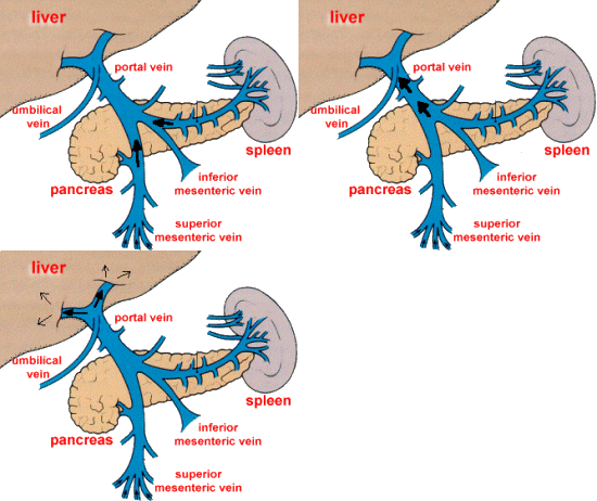
Now the portal vein is something with which you should become very familiar– not just because you see it every time you scan an abdomen, but it is also one of the most commonly ordered vascular Doppler studies ordered for radiology ultrasound–so make it a point to get to know it now and love it later.
To best understand portal venous flow it is first important to understand what is normally occurring in the abdomen so that you understand how common pathology will affect it.
The portal vein is a result of the joining of veins from two areas in the body–the mesenteric veins and the splenic vein —these veins direct all the blood flow from the spleen and bowels into the liver to be cleaned and to harvest nutrients.
So first stop and picture this–blood from the bowels travels upwards/superiorly towards the head of the pancreas, and blood from the spleen travels from left to right towards the pancreatic head where it becomes the portal vein.
So, by this diagram, you should be able to picture how the blood should flow based on where you place your probe. I absolutely always recommend utilizing an intercostal approach to assess the direction of flow–you can almost always visualize the portal vein on anyone despite body habitus/obstacles common to ultrasound. This allows you to become familiar with what you should see every time. RED flow–intercostally, the flow will be advancing towards the transducer so normal should be red. Normal is also called hepatopedal flow—-whereas abnormal is called hepatofugal flow–now you may not be exposed enough to be able to readily identify flow terminology–so here’s the trick I used in school, and still use by the way: hepatofugal is bad–its reversal of flow caused by a resistive force that prevents entry of blood into the liver–hepatofugal is as bad as the f-word. We’ll call it ‘fudge’ as a tribute to the movie ” A Christmas Story” that I absolutely love for exactly 28 days commencing immediately post-Thanksgiving day until the 25th of December–no later. I digress. So I think in my head hepatofudgal. I know less than academic, but it works. No ‘fudging’-fail method of remembering the direction of hepatic flow.
Now, when a patient has cirrhosis, the liver becomes tough like a bad steak that’s hard to get your teeth through–same for blood through the liver–the blood doesn’t want to slow its roll, so it keeps on coming, but has nowhere to go, so it backs up into the vessels it came from.. Think of a panty hose on a faucet that people like my parents used to use to catch lint from the washer–when the hose becomes full of lint, its like a cirrhotic liver–and what happens (I know because I used to watch it) the hose becomes dilated like a water balloon as the water backs up, and then it comes out of whatever place gives the least resistance.. So in the case of cirrhosis, it will back up into the portal vein first–and you’ll notice its dilated (2 cm is upper of norm) when you turn on the color, it will be EITHER blue, OR bidirectional–(bidirectional is a turbulent mix of red and blue and indicates early fibrotic changes–and it will reflect in the waveform as well.)
- Turbulence in Portal Vein
And then of course, you will look at other areas that will hold the overflow, refer back to the picture, where will it back up into?

Splenic varices with PHTN
Spleen-it will be big and bloated with blood–bigger than 12 cm and typically have varices from the increased pressure and volume of blood backing up from the liver


Umbilical vein (remnant reopens under pressure) you’ll see it as a weird vessel smack dab in the middle of the left lobe of the liver.
And now you got it, right?


You must be logged in to post a comment.